General description
CD44 antigen (UniProt: P16070; also known as CDw44, Epican, Extracellular matrix receptor III, ECMR-III, GP90 lymphocyte homing/adhesion receptor, HUTCH-I, Heparan sulfate proteoglycan, Hermes antigen, Hyaluronate receptor, Phagocytic glycoprotein 1, PGP-1, Phagocytic glycoprotein I, PGP-I) is encoded by the CD44 (also known as LHR, MDU2, MDU3, MIC4) gene (Gene ID: 960) in human. CD44 is a single-pass type I membrane glycoprotein that is involved in cell-cell interactions, cell adhesion, and migration. It cell-cell and cell-matrix interactions are mediated through its affinity for hyaluronic acid (HA) and through other ligands. Adhesion with HA is considered to be important in cell migration, tumor growth, and progression. In cancer cells, it may play an important role in the formation of invadopodia. CD44 is synthesized with a signal peptide (aa 1-20), which is subsequently cleaved off to produce mature form. Its extracellular domain is localized to amino acids 21-649 and it has a short helical transmembrane domain (aa 650-670), and a cytoplasmic tail (aa 671-742). Nineteen different isoforms of CD44 are reported that are produced by alternative splicing.
Specificity
Clone hermes-1 is a rat monoclonal antibody that detects human CD44.
Immunogen
Human tonsil lymphocytes.
Application
Anti-CD44, clone Hermes-1, Cat. No. MABF2092, is a rat monoclonal antibody that detects human CD44 and has been tested for use in Flow Cytometry, Immunohistochemistry (Paraffin), and Immunoprecipitation.
Immunohistochemistry Analysis: A representative lot detected CD44 in Immunohistochemistry applications (Jalkanen, S.T., et. al. (1986). Eur J Immunol. 16(10):1195-202).
Immunoprecipitation Analysis: A representative lot immunoprecipitated CD44 in Immunoprecipitation applications (Jalkanen, S.T., et. al. (1986). Eur J Immunol. 16(10):1195-202; Jalkanen, S., et. al. (1987). J Cell Biol. 105(2):983-90).
Flow Cytometry Analysis: A representative lot detected CD44 in Flow Cytometry applications (Jalkanen, S.T., et. al. (1986). Eur J Immunol. 16(10):1195-202).
Research Category
Inflammation & Immunology
Quality
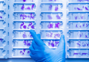
Evaluated by Immunohistochemistry in human skin and human sweat gland tissues.
Immunohistochemistry Analysis: A 1:50 dilution of this antibody detected CD44 in human skin and human sweat gland tissues.
Target description
81.54 kDa calculated for isoform 1.
Physical form
Format: Purified
Protein G purified
Purified rat monoclonal antibody IgG2a in PBS without azide.
Storage and Stability
Stable for 1 year at -20°C from date of receipt.
Handling Recommendations: Upon receipt and prior to removing the cap, centrifuge the vial and gently mix the solution. Aliquot into microcentrifuge tubes and store at -20°C. Avoid repeated freeze/thaw cycles, which may damage IgG and affect product performance.
Other Notes
Concentration: Please refer to lot specific datasheet.
Disclaimer
Unless otherwise stated in our catalog or other company documentation accompanying the product(s), our products are intended for research use only and are not to be used for any other purpose, which includes but is not limited to, unauthorized commercial uses, in vitro diagnostic uses, ex vivo or in vivo therapeutic uses or any type of consumption or application to humans or animals.
- UPC:
- 51202002
- Condition:
- New
- Availability:
- 3-5 Days
- Weight:
- 1.00 Ounces
- HazmatClass:
- No
- MPN:
- MABF2092-100UG


