General description
Type III collagen (also known as COL3A1), which adds structure and strength to connective tissues, is found in many places in the body, especially skin, lung, intestinal walls, and the walls of blood vessels. Collagen type III is initially produced as pro-collagen, a protein consisting of three pro-alpha1(III) chains that form the triple-stranded, rope-like molecule. After being synthesized, the pro-collagen molecule is modified by the cell. Enzymes modify the amino acids lysine and proline in the protein strands by adding chemical groups that are necessary for the strands to form a stable molecule and then later to crosslink to other molecules outside the cell. Other enzymes add sugars to the protein. The type III pro-collagen molecules are released from the cell and are processed by enzymes that clip small segments off either end of the molecules to form mature collagen. The mature collagen molecules assemble into fibrils. Cross-linking between molecules produces a very stable fibril, contributing to collagen′s tissue strengthening function.
Specificity
This antibody detects collagen type III. There is no evidence for cross reactivity with Collagen Types I, V and VI or connective tissue proteins (Elastin, Fibronectin and Laminin) at suggested working concentrations.
Immunogen
Epitope: N-terminus
Human type III collagen (Werkmeister, J.A., et al. 1990).
Application
ELISA Analysis: A previous lot of this antibody was used in ELISA (Werkmeister, J.A., et al., 1991).
Western Blot Analysis: A previous lot of this antibody was used to detect collagen type III in western blot under non-reduced conditions (Werkmeister J.A., et al., 1988; Ramshaw, J.S., et al., 1988).
Some Collagen samples can be contaminated with other Collagen Types. When purified Collagen is used in an application the purity of the Collagen sample should be verified by SDS-page to minimize the risk of false positives.
Immunohistochemistry Analysis: A previous lot of this antibody was used to detect collagen type III in immunohistochemistry (Werkmeister J.A., et al., 1989; Werkmeister J.A., et al., 1989; Werkmeister J.A., et al., 1988).
Research Category
Cell Structure
Research Sub Category
ECM Proteins
This Anti-Collagen Type III Antibody, clone IE7-D7 is validated for use in ELISA, WB, IH for the detection of Collagen Type III.
Quality
Evaluated by Immunohistochemistry in rat knee joint tissue.
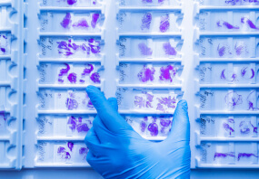
Immunohistochemistry Analysis: A 1:600 dilution of this antibody detected Collagen Type III in rat knee joint tissue.
Target description
138 kDa calculated
Physical form
Format: Purified
Protein G Purified
Purified mouse monoclonal IgG1κ in buffer containing 0.1 M Tris-Glycine (pH 7.4), 150 mM NaCl with 0.05% sodium azide.
Storage and Stability
Stable for 1 year at 2-8°C from date of receipt.
Analysis Note
Control
Rat knee joint tissue
Other Notes
Concentration: Please refer to the Certificate of Analysis for the lot-specific concentration.
This clone displays a high affinity for human, dog, rat, kangaroo and porcine Type III Collagens.
Disclaimer
Unless otherwise stated in our catalog or other company documentation accompanying the product(s), our products are intended for research use only and are not to be used for any other purpose, which includes but is not limited to, unauthorized commercial uses, in vitro diagnostic uses, ex vivo or in vivo therapeutic uses or any type of consumption or application to humans or animals.
Shipping Information:
Dry Ice Surcharge & Ice Pack Shipments: $40
More Information: https://cenmed.com/shipping-returns
- UPC:
- 51172415
- Condition:
- New
- Availability:
- 3-5 Days
- Weight:
- 1.00 Ounces
- HazmatClass:
- No
- MPN:
- MAB3392
- Temperature Control Device:
- Yes


