General description
LAT (linker for activation of T cells) is a leukocyte type III transmembrane adaptor protein. It is made of 262 amino acids, and has a shorter isoform made of 233 amino acids. It is a lipid raft associated protein, and does so by its two cysteine residues present proximal to the membrane. This protein resides in the plasma membrane as well as intracellular and subsynaptic vesicles. It has a small exoplasmic region, a single membrane spanning domain, and a cytoplasmic region containing nine tyrosine residues. This gene maps to human chromosome 16p11.2, spans 5.7kb and has 11 exons.
Immunogen
Linker for activation of T-cells family member 1 recombinant protein epitope signature tag (PrEST)
Application
All Prestige Antibodies Powered by Atlas Antibodies are developed and validated by the Human Protein Atlas (HPA) project and as a result, are supported by the most extensive characterization in the industry.
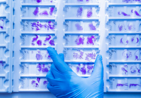
The Human Protein Atlas project can be subdivided into three efforts: Human Tissue Atlas, Cancer Atlas, and Human Cell Atlas. The antibodies that have been generated in support of the Tissue and Cancer Atlas projects have been tested by immunohistochemistry against hundreds of normal and disease tissues and through the recent efforts of the Human Cell Atlas project, many have been characterized by immunofluorescence to map the human proteome not only at the tissue level but now at the subcellular level. These images and the collection of this vast data set can be viewed on the Human Protein Atlas (HPA) site by clicking on the Image Gallery link. We also provide Prestige Antibodies® protocols and other useful information.
Biochem/physiol Actions
LAT (linker for activation of T cells) plays an essential role in the development and activation of T-cells, and T-cell receptor (TCR) signaling. Phosphorylated Zap-70 (Zeta-chain-associated protein kinase 70) phosphorylates the tyrosine residues of LAT. This activated LAT then initiates TCR-signaling. Activated LAT induces multiple signaling intermediates, leading to Ca2+ influx, activation of NFAT (nuclear factor of activated T cells), which eventually leads to the activation of cytokines. It regulates the proliferation of lymphocytes, and controls the differentiation of T-cells into T-helper 1 (Th1) and Th2 subtypes. Activation of natural killer (NK) cells, lead to the phosphorylation and activation of LAT, which in turn interacts with various phosphotyrosine containing proteins, such as PCLγ (phospholipase Cγ). This leads to an overall increase in the cytotoxic function of NK cells. LAT is overexpressed in severe aplastic anemia (SAA), where up-regulated LAT might lead to increased activity of T-cells, and disruption of Th1/Th2 balance.
Features and Benefits
Prestige Antibodies® are highly characterized and extensively validated antibodies with the added benefit of all available characterization data for each target being accessible via the Human Protein Atlas portal linked just below the product name at the top of this page. The uniqueness and low cross-reactivity of the Prestige Antibodies® to other proteins are due to a thorough selection of antigen regions, affinity purification, and stringent selection. Prestige antigen controls are available for every corresponding Prestige Antibody and can be found in the linkage section.
Every Prestige Antibody is tested in the following ways:
- IHC tissue array of 44 normal human tissues and 20 of the most common cancer type tissues.
- Protein array of 364 human recombinant protein fragments.
Linkage
Corresponding Antigen APREST72225.
Physical form
Solution in phosphate-buffered saline, pH 7.2, containing 40% glycerol and 0.02% sodium azide
Legal Information
Prestige Antibodies is a registered trademark of Sigma-Aldrich Co. LLC
- UPC:
- 41116127
- Condition:
- New
- Weight:
- 1.00 Ounces
- HazmatClass:
- No
- WeightUOM:
- LB
- MPN:
- HPA011157-100UL


