General description
E3 ubiquitin-protein ligase Midline-1 (UniProt: O15344; also known as EC: 2.3.2.27, Midin, Putative transcription factor XPRF, RING finger protein 59, RING finger protein Midline-1, RING-type E3 ubiquitin transferase Midline-1, Tripartite motif-containing protein 18) is encoded by the MID1 (also known as FXY, RNF59, TRIM18, XPRF) gene (Gene ID: 4281) in human. Midlin is homodimeric protein with E3 ubiquitin ligase activity towards IGBP1, promoting its monoubiquitination that results in deprotection of the catalytic subunit of protein phosphatase PP2A and its subsequent degradation by polyubiquitination. It can also form heterodimer with MID2. It associates with microtubules throughout the cell cycle, co-localizing with cytoplasmic fibers in interphase and with the mitotic spindle and midbodies during mitosis and cytokinesis. It is highly expressed in fetal kidney, followed by brain and lung. Lower levels are observed in fetal liver. In the adult, it is most abundant in heart, placenta, and brain. Midlin has multiple zinc binding sites and possesses three zinc fingers (one RING type and two B box-type). Mutations in MID1 gene are reported to cause Opitz GBBB syndrome 1 that is characterized by reduced affinity for microtubules, hypertelorism, and genital-urinary defects.
Specificity
This rabbit polyclonal antibody detects E3 ubiquitin-protein ligase Midline-1 (MID1) in Human cells. It targets an epitope within 13 amino acids from the N-terminal region.
Immunogen
Epitope: N-terminus
KLH-conjugated linear peptide corresponding to 13 amino acids from the N-terminal region of human E3 ubiquitin-protein ligase Midline-1.
Application
Anti-MID1, Cat. No. ABN1374, is a highly specific rabbit polyclonal antibody that targets E3 ubiquitin-protein ligase Midline-1 and has been tested for use in Immunohistochemistry (Paraffin) and Western Blotting.
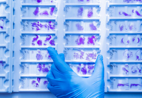
Immunohistochemistry Analysis: A 1:250 dilution from a representative lot detected MID1 in human placenta and human cerebral cortex tissue sections.
Research Category
Neuroscience
Quality
Evaluated by Western Blotting in HeLa cell lysate.
Western Blotting Analysis: 1 µg/mL of this antibody detected MID1 in HeLa cell lysate.
Target description
~75 kDa observed; 75.25 kDa calculated.
Physical form
Affinity Purified
Format: Purified
Purified rabbit polyclonal antibody in buffer containing 0.1 M Tris-Glycine (pH 7.4), 150 mM NaCl with 0.05% sodium azide.
Storage and Stability
Stable for 1 year at 2-8°C from date of receipt.
Other Notes
Concentration: Please refer to lot specific datasheet.
Disclaimer
Unless otherwise stated in our catalog or other company documentation accompanying the product(s), our products are intended for research use only and are not to be used for any other purpose, which includes but is not limited to, unauthorized commercial uses, in vitro diagnostic uses, ex vivo or in vivo therapeutic uses or any type of consumption or application to humans or animals.
biological source: rabbit. Quality Level: 100. antibody form: affinity isolated antibody. antibody product type: primary antibodies. clone: polyclonal. species reactivity: human. species reactivity (predicted by homology): bovine (based on 100% sequence homology), rat (based on 100% sequence homology), mouse (based on 100% sequence homology). packaging: antibody small pack of 25 . μ. g. technique(s): immunohistochemistry: suitable (paraffin), western blot: suitable. NCBI accession no.: NP_000372.1. UniProt accession no.: O15344. target post-translational modification: unmodified. Gene Information: human ... MID1(4281). Storage Class Code: 12 - Non Combustible Liquids. WGK: WGK 1. Flash Point(F): Not applicable. Flash Point(C): Not applicable.- UPC:
- 51342709
- Condition:
- New
- Availability:
- 3-5 Days
- Weight:
- 1.00 Ounces
- HazmatClass:
- No
- MPN:
- ABN1374-100UG


