General description
Prions are thought to cause a number of diseases in a variety of mammals, including bovine spongiform encephalopathy (BSE, also known as "mad cow disease") in cattle and the Creutzfeldt-Jakob disease (CJD) in humans. All thus-far hypothesized prion diseases affect the structure of the brain or other neural tissue, and all are currently untreatable and thought to be fatal. Prions are hypothesized to infect and propagate by refolding abnormally into a structure which is able to convert normal molecules of the protein into the abnormally structured form. All known prions induce the formation of an amyloid fold, in which the protein polymerises into an aggregate consisting of tightly packed beta sheets. This altered structure is extremely stable and accumulates in infected tissue, causing cell death and tissue damage. This stability means that prions are resistant to denaturation by chemical and physical agents, making disposal and containment of these particles difficult.
Specificity
Prion protein, amino acid residues 109-112 of human, hamster and feline. Does not react with PrP from any other mammalian species. MAB1562 is reactive to native and denatured forms of PrP. Tissue or cells which have been fixed requires that the epitope be re-exposed (see below). Recognizes both protease sensitive and protease resistant forms of PrP.
Immunogen
Epitope: a.a. 109-112
Application
Immunohistochemistry(paraffin):
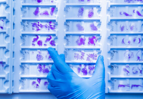
Representative images from a previous lot. Optimal Staining With Citrate Buffer, pH 6.0, Epitope Retrieval: Human Brain
Immunohistochemistry (Kitamoto et al., 1987):
1:100-1:1,000 *See protocol below.
Epitope must be re-exposed in fixed tissue by pretreatment of tissue using one of the following procedures:
a. formic acid for 10 minutes at room temperature (Kitamoto et al., 1987)
b. hydrolytic autoclaving (Kitamoto et al., 1991)
c. microwaving (BioGenex, San Ramon, CA)
Western Blot: (Kascsak, R.J., 1991; Kascsak, R.J., 1987):
1:10,000-1:100,000 dilution of a previous lot was used.
Immunoprecipitation: (Kascsak, R.J., 1991; Kascsak, R.J., 1987):
1:10-1:100 dilution of a previous lot was used.
ELISA: (Kascsak, R.J., 1991; Kascsak, R.J., 1987):
Optimal working dilutions must be determined by end user.
This Anti-Prion Protein Antibody, a.a. 109-112, clone 3F4 is validated for use in ELISA, IH, IH(P), IP, WB for the detection of Prion Protein.
Quality
Immunohistochemistry(paraffin):
Prion Protein (cat. # MAB1562) staining pattern/morphology in normal brain. Tissue was pretreated with Citrate, pH 6.0. This lot of antibody was diluted to 1:500, using IHC-Select
Optimal Staining With Citrate Buffer, pH 6.0, Epitope Retrieval: Human Brain
Target description
12.3 kDa
Physical form
Format: Purified
Purified mouse monoclonal IgG2a in buffer containing PBS and no preservative.
Legal Information
CHEMICON is a registered trademark of Merck KGaA, Darmstadt, Germany
Shipping Information:
Dry Ice Surcharge & Ice Pack Shipments: $40
More Information: https://cenmed.com/shipping-returns
- UPC:
- 51172419
- Condition:
- New
- Availability:
- 3-5 Days
- Weight:
- 1.00 Ounces
- HazmatClass:
- No
- MPN:
- MAB1562
- Temperature Control Device:
- Yes


