General description
Major prion protein (UniProt P04156; also known as ASCR, CD230, PrP, PrP27-30, PrP33-35C) is encoded by the PRNP (also known as ALTPRP, CJD, GSD, HDL1, KURU, PRIP, PRP, SENF) gene (Gene ID 5621) in human. The major prion protein (PrP) is synthesized with an N-terminal signal peptide (a.a. 1-22) and a C-terminal proptptide (a.a. 231-253) sequence, which are posttranslationally removed to yield the mature (a.a. 23-230) N-glycosylated, glycosylphosphatidylinositol (GPI)-anchored cell surface protein. Structurally, PrP consists of a long, disordered, flexible NH2-proximal region and a globular COOH-proximal domain linked together by a hydrophobic core (HC) region (a,a. 111-134) that is considered as a key region in both physiological and disease related processes involving the prion protein. The N-terminal region is composed of five octapeptide (PHGGGWGQ) repeats (a.a. 51-91) sandwiched between two positively charged clusters, CC1 & CC2 (a.a. 23-27 & 95-110). The exact physiological function of PrP is not known, although various biological functions have been suggested for this protein, including signal transduction, neurotransmitter metabolism, cell adhesion, antioxidant activity, neurogenesis, immune cell activation, copper metabolism and homeostasis of trace elements. Transmissible spongiform encephalopathies (TSEs) represent a family of rare and fatal neurodegenerative disorders, including Creutzfeldt-Jakob disease, Gerstmann-Sträussler-Scheinker syndrome, fatal familial insomnia and kuru in human, bovine spongiform encephalopathy in cattle, and scrapie in sheep. It is widely accepted that the conformational transition of the native and predominantly α-helical cellular PrP (PrPC) to a β-sheet-rich pathogenic scrapie isoform (PrPSc) is responsible for the accumulation of PrPSc aggregates.
Specificity
Clone 12D6.1 detects an epitope present in human PrP and the spliced isoform PrP(M8) reported by UniProt (P04156-1 & P04156-2), but absent in the alternative prion protein AltPrP (UniProt F7VJQ1-1).
Immunogen
Epitope: Internal (N-terminal half).
Human IgG1 Fc-tagged recombinant human prion protein internal fragment.
Application
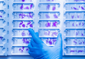
Immunohistochemistry Analysis: A 1:50 dilution from a representative lot detected prion protein in human cerebral cortex tissue sections.
Research Category
Neuroscience
Research Sub Category
Neurodegenerative Diseases
This Anti-Prion Protein Antibody, clone 12D6.1 is validated for use in Western Blotting, Immunohistochemistry (Paraffin) for the detection of Prion Protein.
Quality
Evaluated by Western Blotting of recombinant human prion protein.
Western Blotting Analysis: 1.0 µg/mL of this antibody detected 0.05 µg of human IgG1 Fc-tagged recombinant human prion protein internal fragment.
Target description
27.66/26.89 kDa (PrP/PrP(M8) prepro-form), 22.75/25.24 kDa (mature/pro-form) calculated.
Linkage
Replaces: MAB1562
Physical form
Format: Purified
Protein G purified.
Purified mouse monoclonal IgG2aκ antibody in buffer containing 0.1 M Tris-Glycine (pH 7.4), 150 mM NaCl with 0.05% sodium azide.
Storage and Stability
Stable for 1 year at 2-8°C from date of receipt.
Other Notes
Concentration: Please refer to lot specific datasheet.
Disclaimer
Unless otherwise stated in our catalog or other company documentation accompanying the product(s), our products are intended for research use only and are not to be used for any other purpose, which includes but is not limited to, unauthorized commercial uses, in vitro diagnostic uses, ex vivo or in vivo therapeutic uses or any type of consumption or application to humans or animals.
Shipping Information:
Dry Ice Surcharge & Ice Pack Shipments: $40
More Information: https://cenmed.com/shipping-returns
- UPC:
- 51183619
- Condition:
- New
- Availability:
- 3-5 Days
- Weight:
- 1.00 Ounces
- HazmatClass:
- No
- MPN:
- MABN1186
- Temperature Control Device:
- Yes


